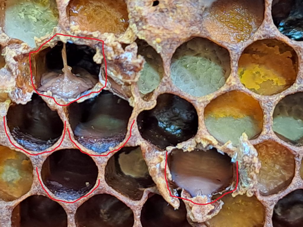
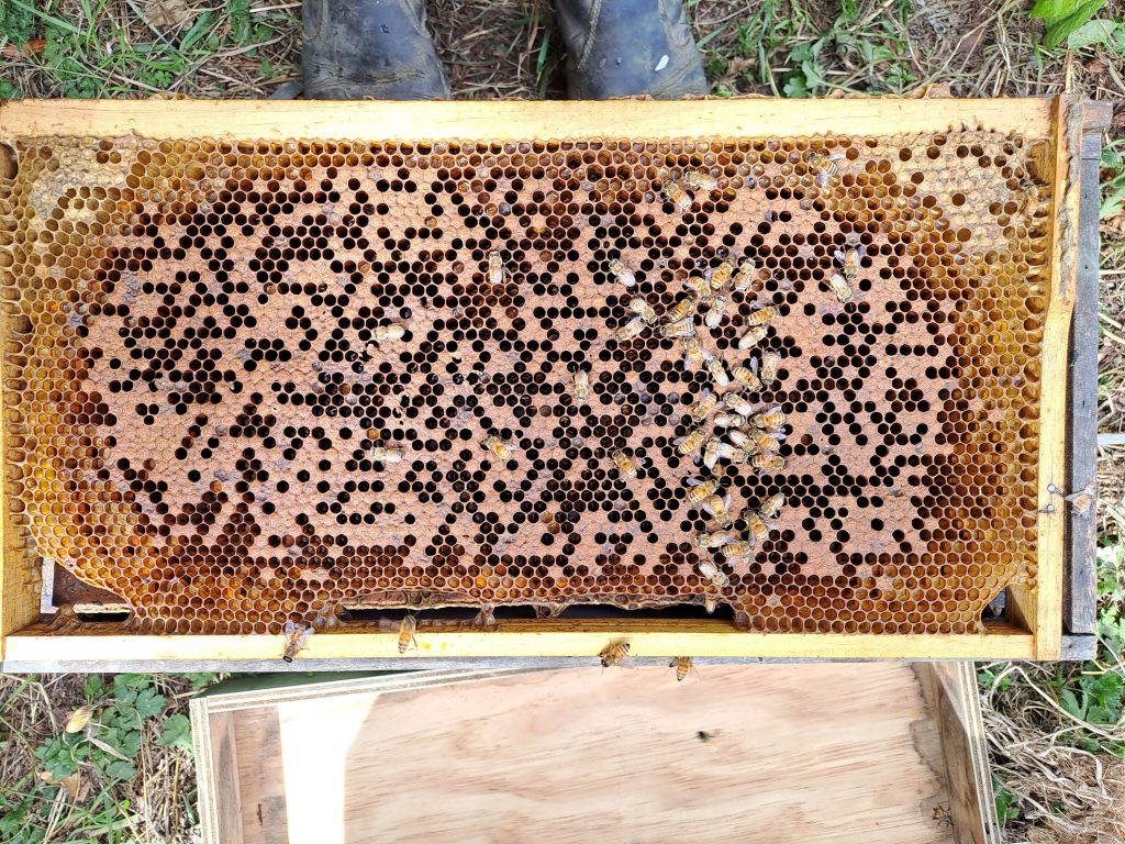
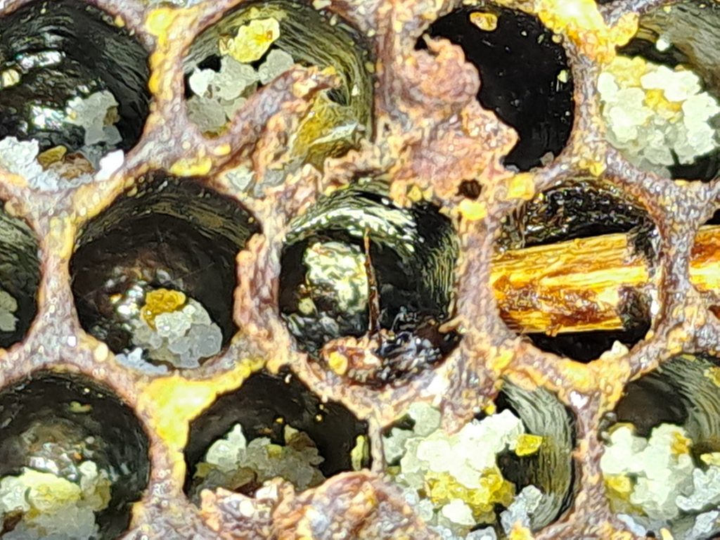
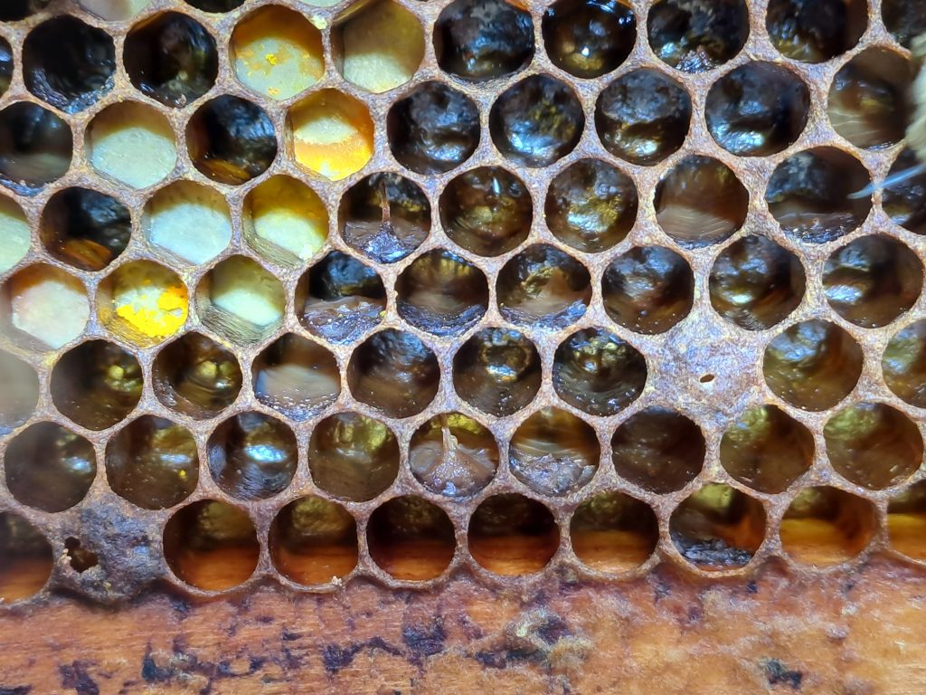
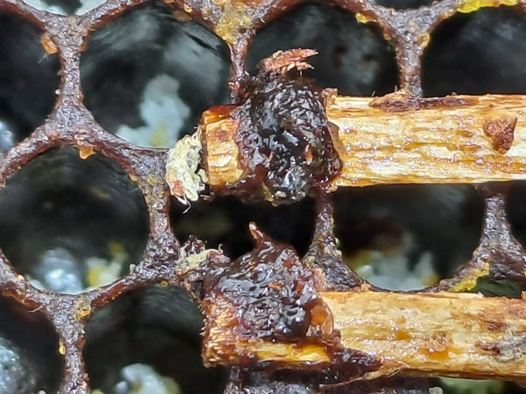
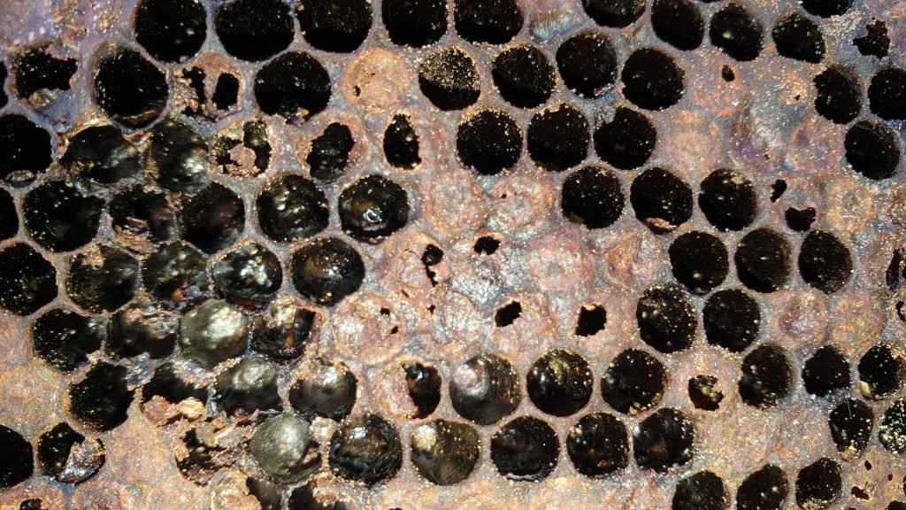
AFB Photo Gallery
We invite all beekeepers to send in their photos of AFB and the destruction of their hives in order to help educate other beekeepers. Please send your photos to [email protected] with a brief description of each shot. For best results, the photos need to be in high resolution.
Clicking on an image will open it in a new window and a larger size.


AFB roping out – video courtesy of Dan Childs.
AFB roping out – video courtesy of Tom Hurford


Video courtesy of Blake Cole
Take the AFB 5 minute quiz
How well do you know what you need to know about AFB and beekeeping? Take our short quiz and find out.
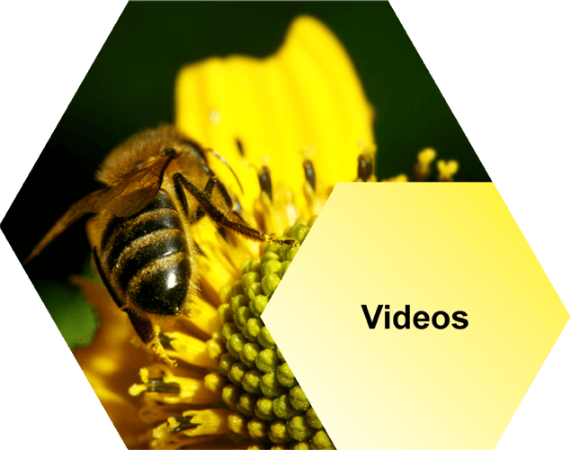
Videos
Our videos cover everything from your legal obligations to how to recognise AFB, collecting cell and bee samples and more.
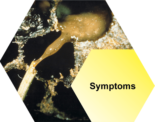
Symptoms
There’s a lot of good information here, telling you everything you need to know about recognising AFB: the visual symptoms, smell of AFB and more.
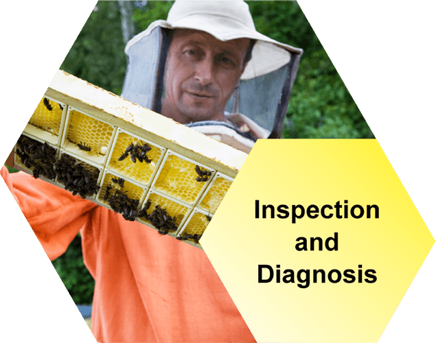
Inspection and Diagnosis
Successfully eliminate AFB by telling the difference between symptoms of AFB and other brood diseases in the hive. We tell you the best methods for inspecting your hives.
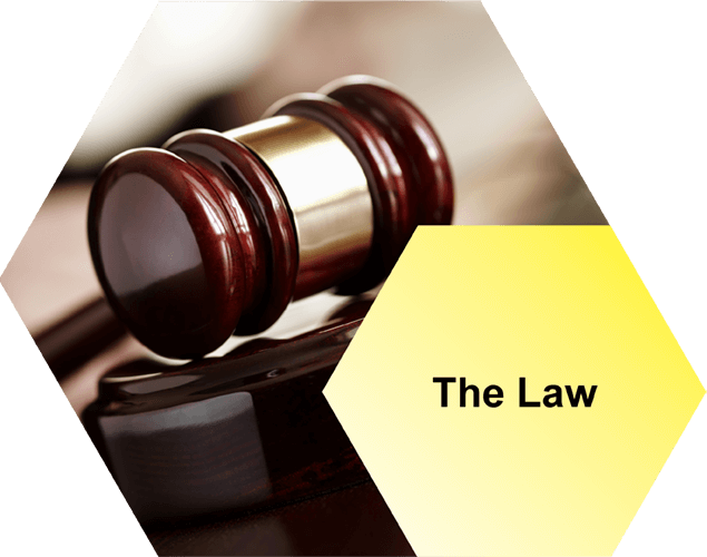
The Law
New Zealand beekeepers have a number of legal obligations that must be met regarding AFB disease. Read the shortened list in summary, here.
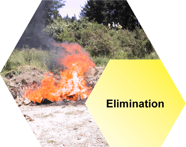
Elimination
Most hives become infected because bees, honey or equipment have been put into a hive from another hive that is infected with AFB. Lower your chances of an AFB infection by reading this section.
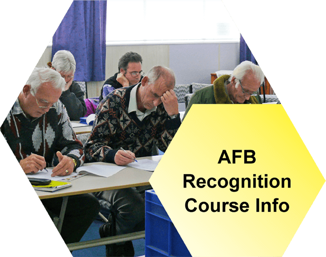
AFB Recognition Course Info
Find out when the next AFB Recognition and Competency Courses, or Refresher Courses are available. These are held throughout the year in various New Zealand locations across the South Island and North Island.
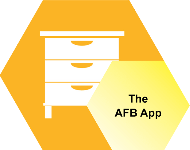
The AFB App
Follow the link below to open the App. Once open to save to your device you need to bookmark the URL on your phone so you can find it easily again. Please click here to open.